Agriculture is experiencing a paradigm shift through the integration of advanced imaging technologies. Among these innovations, thermal imaging stands out as a powerful tool for monitoring crop health and detecting diseases at their inception. By capturing the subtle differences in temperature across plant canopies, farmers and agronomists can gain critical insights into plant physiology and stress responses. The following sections explore the foundational concepts, practical applications, and future directions of thermal imaging in crop disease detection.
Principles of Thermal Imaging
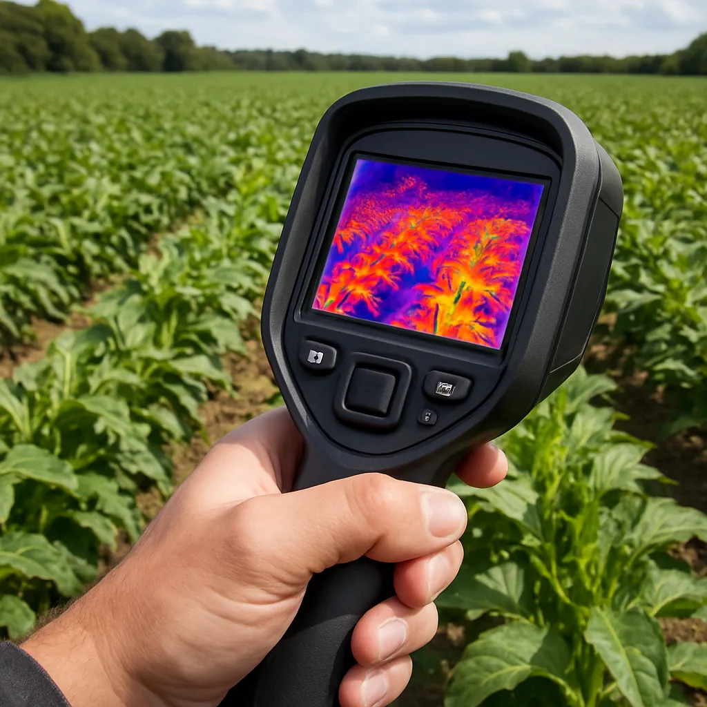
Thermal imaging harnesses the power of infrared radiation to visualize temperature variations across surfaces. Every object emits radiation proportional to its temperature, and specialized cameras convert this signal into a temperature map. In an agricultural setting, subtle thermal anomalies often correspond to physiological changes in plants. For instance, water-stressed or diseased crops may exhibit higher surface temperatures due to reduced transpiration rates.
Key elements of thermal imaging systems include:
- Sensors with high sensitivity to detect minute temperature differences.
- Calibration routines to correct for environmental factors such as wind or ambient temperature.
- Algorithms for processing raw thermal data into actionable maps.
The spatial resolution and sensitivity of the camera define its ability to spot early signs of stress. Higher-resolution devices reveal finer details within the canopy, enabling the identification of small clusters of affected plants. Combined with robust data processing, thermal imaging facilitates early intervention and precise management strategies.
Implementing Thermal Imaging for Disease Detection
Integrating thermal imaging into crop management involves multiple steps, from data acquisition to interpretation:
- Platform Selection: Ground-based units, tractor-mounted cameras, or drone-based thermal systems.
- Flight Planning: For aerial surveys, flight altitude, speed, and overlap dictate image coverage and quality.
- Time of Day: Optimal image capture often occurs under stable thermal conditions, typically during early morning or late afternoon.
- Data Processing: Specialized algorithms transform thermal frames into georeferenced maps, highlighting hotspots and cold zones.
When a pathogen invades a plant, physiological changes such as reduced water transport or cell damage alter its thermal signature. For example, fungal infections often manifest as localized hotter regions on leaves, while viral diseases may cause cooler spots due to impaired metabolism. By comparing baseline thermal profiles with current images, agronomists can pinpoint affected areas before symptoms become visible to the naked eye.
Successful implementation has been demonstrated in multiple crops:
- Vineyards: Detecting powdery mildew outbreaks through canopy temperature shifts.
- Cereal Fields: Mapping drought-stressed areas that are prone to Fusarium infection.
- Horticulture: Monitoring greenhouse tomatoes for early bacterial and viral diseases.
These examples illustrate the potential of thermal imaging to enhance precision agriculture practices, reduce chemical inputs, and promote crop sustainability.
Case Study: Thermal Signatures in Wheat
In a field trial, researchers equipped a tractor-mounted thermal camera to survey a wheat field infected with stripe rust. Over successive passes, they observed:
- Distinct thermal anomalies that preceded visible rust pustules by several days.
- A correlation between infection severity and canopy temperature increase.
- Improved fungicide application efficacy when guided by thermal maps.
By overlaying thermal data with multispectral imagery and soil moisture readings, the team developed an integrated disease management plan. The approach led to a 25% reduction in fungicide use and a 15% yield increase compared to conventional scouting methods. This case underscores the capacity of thermal imaging to deliver both environmental and economic benefits.
Challenges and Future Prospects
Despite its promise, thermal imaging faces several hurdles in mainstream adoption:
- Environmental Interference: Fluctuating weather conditions can introduce noise into thermal readings.
- Data Volume: High-resolution surveys generate large datasets, requiring robust storage and processing capabilities.
- Interpretation Complexity: Differentiating between stressors—such as nutrient deficiency versus disease—can be challenging without complementary data.
- Cost: Advanced thermal cameras and UAV platforms involve substantial initial investment.
Ongoing research aims to overcome these barriers by:
- Developing machine-learning models that integrate thermal, spectral, and meteorological data for more accurate diagnosis.
- Innovating low-cost sensor designs to broaden access among smallholder farmers.
- Automating real-time alerts through edge computing on field-deployed devices.
- Exploring synergies with Internet of Things (IoT) networks to deliver continuous monitoring.
As these advances unfold, thermal imaging is poised to transform disease surveillance in agriculture. By enabling rapid detection and targeted response, this technology supports healthier crops, improved resource efficiency, and greater resilience against emerging threats.